
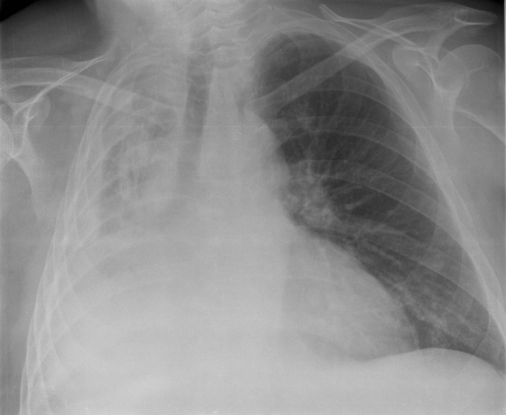
Atelectasis is a medical condition characterized by the partial or complete collapse of a lung or a portion of a lung. It occurs when the tiny air sacs, known as alveoli, within the lung become deflated, resulting in decreased oxygen exchange and impaired lung function.
Types:
There are several types of atelectasis, including:
1. Resorptive Atelectasis: This type occurs when the airway leading to the alveoli becomes blocked, preventing air from entering the affected area of the lung. Common causes include mucus plugs, tumors, or foreign objects obstructing the airway.
2. Compression Atelectasis: Compression of the lung can occur due to external pressure on the chest cavity, such as from a pleural effusion (accumulation of fluid in the pleural space), pneumothorax (collapsed lung due to air leakage into the pleural space), or a tumor pressing on the lung tissue.
3. Contraction Atelectasis: This type is often associated with scarring or fibrosis of the lung tissue, which causes the lung to shrink and pull away from the chest wall.
Signs and Symptoms:
The signs and symptoms of atelectasis vary depending on the extent and location of lung collapse. Common symptoms include shortness of breath, rapid breathing, chest pain, coughing, and decreased oxygen levels in the blood. In severe cases, atelectasis can lead to respiratory failure and life-threatening complications.
Cause or Causative Agents:
Atelectasis can be caused by a variety of factors, including:
- Surgery: Anesthesia and prolonged bed rest after surgery can contribute to shallow breathing and decreased lung expansion, increasing the risk of atelectasis.
- Lung Disorders: Conditions such as pneumonia, bronchitis, asthma, or chronic obstructive pulmonary disease (COPD) can lead to airway inflammation and mucus buildup, which may block the air passages and cause lung collapse.
- Trauma: Injuries to the chest or head can result in pneumothorax or hemothorax, leading to lung collapse.
- Infections: Respiratory infections, especially in infants and young children, can cause mucus buildup and blockage of the airways, leading to atelectasis.
Prevention:
Preventing atelectasis involves maintaining good respiratory hygiene and addressing underlying conditions that may increase the risk of lung collapse. Measures such as deep breathing exercises, coughing techniques, early mobilization after surgery, and smoking cessation can help prevent atelectasis.
Control:
Treatment for atelectasis aims to re-expand the collapsed lung tissue and improve ventilation. Depending on the underlying cause and severity of symptoms, interventions may include:
- Incentive Spirometry: Breathing exercises using a device called a spirometer can help improve lung function and prevent atelectasis after surgery.
- Chest Physiotherapy: Techniques such as percussion, vibration, and postural drainage can help loosen and remove mucus from the airways, allowing for better airflow and lung expansion.
- Bronchoscopy: This procedure may be performed to remove mucus plugs, foreign objects, or tumors blocking the airways.
- Oxygen Therapy: Supplemental oxygen may be administered to improve oxygenation and alleviate symptoms of hypoxemia (low blood oxygen levels).
In conclusion, atelectasis is a common respiratory condition characterized by lung collapse due to various factors. Early recognition, prevention, and appropriate management are essential to prevent complications and improve outcomes for individuals affected by this condition.